
Tomography is the process of generating a 2-dimensional image of a slice or section through a 3-dimensional object. Similar to looking at one slice of bread within the whole loaf.
- A CT scan uses data from several X-ray images of structures inside the body and converts them into pictures.
- The technique utilizes digital geometry processing to generate 3D images.
- CT scans are a source of ionizing radiation and can cause cancer.
- A CT scanner emits a series of narrow beams through the human body, producing more detail than standard single beam X-rays.
- CT scanners are able to distinguish tissues inside a solid organ.
- Contrast dyes are sometimes used to improve the clarity of the image.
- CT scanning is particularly useful for getting detailed 3D images of certain parts of the body, such as soft tissues, blood vessels, and the brain.
- The images created are analyzed by radiologists.
- Unlike MRI scans, a CT scan uses X-rays.
- A CT scan is able to illustrate organ tear and organ injury quickly and so is often used for accident victims.
What is a CT scan?
The CT scanner uses digital geometry processing to generate a 3-dimensional (3D) image of the inside of an object. The 3D image is made after many 2-dimensional (2D) X-ray images are taken around a single axis of rotation – in other words, many pictures of the same area are taken from many angles and then placed together to produce a 3D image.
The Greek word tomos means “slice”, and the Greek word graphein means “write”.
Although CT is a useful tool for assisting diagnosis in medicine, it is a source of ionizing radiation and can cause cancer. The National Cancer Institute1 advises patients to discuss the risks and benefits of computerized tomography with their doctors.
How do CT scans work?
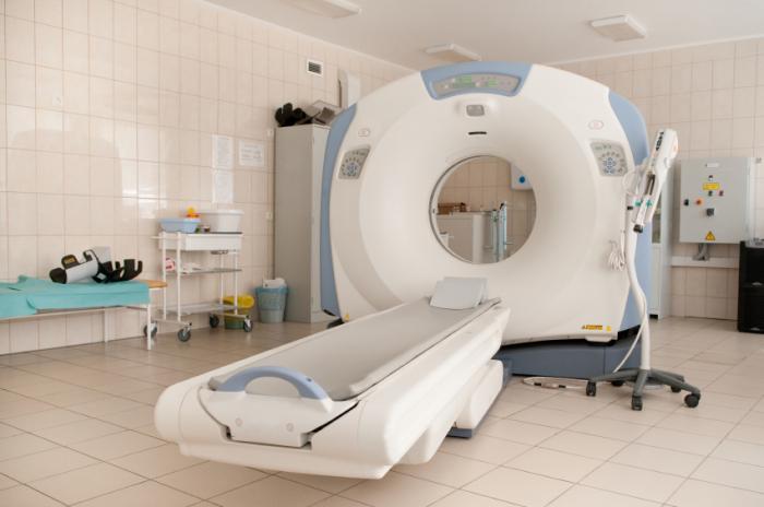
A CT imaging suite.
A CT scanner emits a series of narrow beams through the human body as it moves through an arc, unlike an X-ray machine which sends just one radiation beam. The final picture is far more detailed than an X-ray image.
Inside the CT scanner there is an X-ray detector which can see hundreds of different levels of density. It can see tissues inside a solid organ. This data is transmitted to a computer, which builds up a 3D cross-sectional picture of the part of the body and displays it on the screen.
Sometimes a contrast dye is used because it shows up much more clearly on the screen. If a 3D image of the abdomen is required the patient may have to drink a barium meal. The barium appears white on the scan as it travels through the digestive system. If images lower down the body are required, such as the rectum, the patient may be given a barium enema. If blood vessel images are the target, the barium will be injected.
The accuracy and speed of CT scans may be improved with the application of spiral CT. The X-ray beam takes a spiral path during the scanning – it gathers continuous data with no gaps between images. For a spiral scan of the chest, for example, the patient will be asked to hold his/her breath for a few seconds.
What are they like for patients?
Most places will provide the patient with a gown. He/she will need to undress, usually down to their underwear, and put the gown on. If the place does not provide a gown the patient should wear loose-fitting clothes.
Doctors may ask the patient to fast (eat nothing) and even refrain from consuming liquids for a specific period before the scan.
The patient will be asked to lie down on a motorized examination table, which then goes into the giant doughnut-like machine. The couch with the patient goes into the doughnut hole.
Some patients may be given a contrast dye or substance which is either swallowed, given as an enema, or injected. This improves the picture of some blood vessels or tissues. If a patient is allergic to contrast material he/she should tell the doctor beforehand. There are some medications that reduce allergic reactions to contrast materials.
As metal interferes with the workings of the CT scanner the patient will need to remove all jewelry and metal fastenings. In the majority of cases the patient will lie on his/her back, facing up. But sometimes it may be necessary to lie face-down or sideways.
After the machine has taken one X-ray picture, the couch will move slightly, and then another picture is taken, etc. The patient needs to lie very still for best results.
During the scan everybody except for the patient will leave the room. The radiographer will still be able to communicate with the patient, and vice-versa, through an intercom. If the patient is a child, a parent or adult might be allowed to stand or sit nearby – that person will have to wear a lead apron to prevent radiation exposure.
Pregnancy – any woman who suspects she may be pregnant should tell her doctor beforehand. The UK’s National Health Service2 warns that CT scans are not recommended for pregnant mothers because of the risk that X-rays might harm the unborn baby.
Claustrophobia – patients who suffer from claustrophobia need to tell their doctor or radiographer beforehand, advises Cancer Research UK3. The patient may be given an injection or tablet to calm them down before the scan.
Breastfeeding – the State Government of Victoria4 in Australia says that mothers should avoid breastfeeding their babies for about 24 hours after a CT scan if an iodinated intravenous dye was used. The dye may pass into the breast milk. If you are breastfeeding, tell your doctor beforehand.
When are CT scans used?
CT scanning is useful to get a very detailed 3D image of certain parts of the body, such as soft tissues, the pelvis, blood vessels, the lungs, the brain, abdomen, and bones.
It is often the preferred method of diagnosing many cancers, such as liver, lung, and pancreatic cancers. The image allows a doctor to confirm the presence of a tumor. The tumor’s size can be measured, plus its exact location, as well as to determine how much the tumor has affected nearby tissue.
A scan of the head can provide the doctor with important information about the brain – he/she may want to know whether there is any bleeding, swelling of the arteries, or tumors.
A CT scan will tell the doctor whether the patient has a tumor in his/her abdomen, and whether any internal organs in that area are swollen or inflamed. It will reveal whether there are lacerations of the spleen, kidneys or liver.
As a CT scan can detect abnormal tissue it is a useful device for planning areas for radiotherapy and biopsies.
A CT scan can also provide valuable data on the patient’s vascular condition. Vascular refers to blood flow. Many vascular conditions can lead to stroke, kidney failure, and even death. It can help a doctor assess bone diseases, bone density, and the state of the patient’s spine.
A CT scan can reveal vital data about injuries to the patient’s hands, feet and other skeletal structures – even small bones can be seen clearly, as well as their surrounding tissue.
Who analyzes the image?
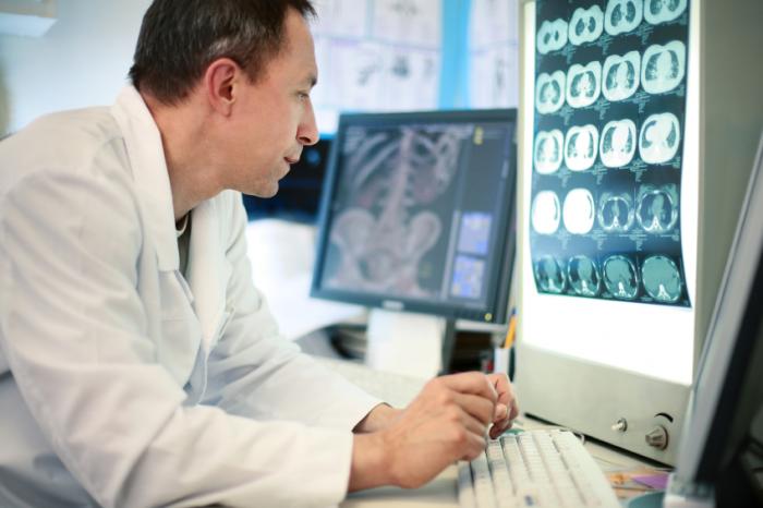
A radiologist who is trained in supervising and interpreting radiology examinations will analyze the images.
A radiologist who is trained in supervising and interpreting radiology examinations will analyze the images and send his/her report to the patient’s doctor. A radiologist is a doctor.
Radiology is a branch of medicine. A radiologist is a fully qualified doctor who specializes in radiology – MRI, CT scans, radiographs, nuclear medicine scans, mammograms and sonograms.
A radiologic technologist or radiographer is the X-ray technician. The person who takes the X-rays.
The radiologist is a doctor; the radiographer is not a doctor.
What are the differences between CT and MRI?
The differences between CT and MRI scans are as follows:
- A CT scan uses X-rays. An MRI does not use X-rays; it uses magnets and radio waves.
- A CT scan does not show tendons and ligaments, an MRI does.
- MRI is better for looking at the spinal cord.
- A CT scan is better for looking at cancer, pneumonia, abnormal chest x-rays, bleeding in the brain (especially from injury).
- A brain tumor is better seen on MRI.
- A CT scan shows organ tear and organ injury more quickly – so it may be the best choice for accident victims.
- Broken bones and vertebrae are better seen on CT scan.
- CT scans are better at visualizing the lungs and organs in the chest cavity between the lungs.